Gallbladder Disease in Dogs: Sludge and Mucoceles

The gallbladder rarely gets a mention in conversations about dog health. It is a small storage sac tucked against the liver whose job is to hold bile between meals and release it into the intestine when fat needs digesting. Most owners never think about it at all — until an ultrasound report comes back with a word they have never heard.
Two findings account for most gallbladder conversations in general practice. The first is biliary sludge, which is extremely common, frequently discovered by accident, and usually of no clinical consequence whatsoever. The second is a gallbladder mucocele, which is far less common but is a genuinely serious condition that can progress to a life-threatening emergency.
The two get confused constantly, partly because they look related on an ultrasound screen and partly because the language sounds similar. Understanding the difference is genuinely useful for owners, because one is usually a note in the file and the other is a reason for careful monitoring and sometimes surgery. This guide explains both, what causes them, how they are found, and what happens next.
Important: This article shares general educational information for pet owners and is not a substitute for an in-person veterinary examination, diagnosis, or treatment. For any medical concern or emergency, contact your veterinarian promptly.
What the Gallbladder Does
Bile is produced continuously by the liver and helps break down and absorb dietary fat. Between meals, bile is diverted into the gallbladder, where it is stored and concentrated. When a fatty meal reaches the small intestine, the gallbladder contracts and releases its contents down the bile duct to do their work.
That storage-and-release cycle depends on the gallbladder contracting properly and on the bile duct remaining open. Problems in this system generally come down to one of three things: bile that has become abnormally thick, a gallbladder that is not emptying well, or an obstruction preventing bile from leaving.
Because bile also carries waste products that the liver has processed for elimination, a blocked biliary system causes those products to back up into the bloodstream. That is why jaundice — a yellow tinge to the gums, the whites of the eyes, and the skin — is one of the classic warning signs of serious gallbladder disease.
- Storage, not production: bile is made by the liver; the gallbladder stores and concentrates it.
- Fat digestion: bile release is triggered largely by fat arriving in the intestine.
- Dogs cope without one: the gallbladder can be removed, and dogs live entirely normal lives afterwards.
- Cats differ: feline gallbladder disease exists but the pattern is different and less common than in dogs.
Biliary Sludge: Usually Nothing
Biliary sludge is thickened, sediment-like material within the gallbladder. On ultrasound it appears as a layer of denser material sitting in the dependent part of the gallbladder, often shifting when the dog changes position.
It is remarkably common, particularly in older dogs, and is frequently found on ultrasounds performed for entirely unrelated reasons. In the great majority of these dogs it causes no signs, no illness, and no consequences. It is what veterinarians call an incidental finding.
Sludge tends to accumulate when the gallbladder is not emptying as frequently or completely as usual — for instance in a dog who has not eaten for a while, or one whose gallbladder motility has been reduced by illness or medication. It can also increase in dogs with certain hormonal conditions or abnormalities of fat metabolism.
The usual approach to sludge in a well dog is straightforward: note it, investigate any concurrent signs on their own merits, and re-check it if the clinical picture changes. Treatment is not typically needed for sludge alone. Where sludge is found alongside abnormal liver values or gastrointestinal signs, your veterinarian will look for the underlying reason rather than assuming the sludge is the culprit.
Gallbladder Mucocele: The Serious One
A gallbladder mucocele is a different proposition. Here the cells lining the gallbladder produce excessive thick mucus, which accumulates until the gallbladder becomes distended with immobile, gelatinous material that will not flow.
On ultrasound this produces a characteristic striated pattern that veterinarians often describe as resembling a kiwi fruit in cross-section. Unlike sludge, this material does not shift when the dog moves, and that lack of movement is one of the features that distinguishes the two.
The problem is progressive. As the gallbladder distends, its wall becomes stretched and its blood supply compromised. The thickened material can obstruct the bile duct, causing bile to back up. In advanced cases the gallbladder wall can rupture, releasing bile into the abdomen and causing a severe, life-threatening peritonitis that requires emergency surgery.
Not every mucocele progresses at the same rate, and some dogs are diagnosed while entirely well and remain stable for a long period under monitoring. But the potential for rapid deterioration is real, which is why a mucocele changes the conversation from observation to active management.
Which Dogs Are at Risk
Gallbladder mucoceles are recognised in many breeds but show clear predispositions.
- Shetland Sheepdogs: the breed most strongly associated with the condition.
- Cocker Spaniels: also notably over-represented.
- Miniature Schnauzers: particularly those with abnormalities of fat metabolism.
- Border Terriers and Pomeranians: among other breeds reported more frequently.
- Middle-aged and older dogs: the condition is uncommon in young animals.
- Dogs with hormonal disease: conditions affecting thyroid or adrenal function are associated.
- Dogs with high blood fat levels: abnormal lipid metabolism is a recognised association.
If your dog belongs to one of the predisposed breeds, it is reasonable to mention that at wellness visits, particularly as your dog reaches middle age and if routine blood work ever shows changes in liver values.
The associations with hormonal and metabolic disease are practically important. A dog diagnosed with a mucocele will usually be screened for these conditions, because identifying and managing an underlying endocrine problem is part of the overall plan.
Signs Owners May Notice
One of the difficulties with gallbladder disease is that many affected dogs show nothing at all until the condition is advanced. When signs do appear, they are frequently vague and could easily be attributed to something minor.
- Vomiting: intermittent at first, sometimes over weeks.
- Reduced appetite: picking at food, or refusing meals that would normally be eaten enthusiastically.
- Lethargy: lower energy, sleeping more, less interest in walks.
- Abdominal discomfort: a hunched posture, reluctance to be picked up, or tension when the belly is touched.
- Jaundice: yellowing of the gums, the whites of the eyes, or the skin inside the ears — a serious sign requiring same-day attention.
- Increased drinking and urinating: often reflecting an associated hormonal condition.
- Diarrhoea: in some dogs.
- Sudden severe illness: collapse, marked pain, and rapid deterioration, which can indicate rupture.
The vague early signs are precisely why gallbladder disease is often found on blood work rather than on the strength of symptoms. A dog who has been slightly off for a week and has raised liver values on a routine panel is a very typical route to diagnosis.
Jaundice deserves special emphasis. Yellow gums or yellow whites of the eyes are never normal in a dog and always warrant urgent veterinary assessment, whatever the underlying cause turns out to be.
How Veterinarians Diagnose It
The pathway usually begins with blood testing. Liver enzyme elevations, particularly those associated with the biliary system, prompt further investigation. Blood work also assesses general health, screens for hormonal disease, and provides baseline values if surgery becomes likely.
Abdominal ultrasound is the key test. It visualises the gallbladder directly, shows whether the contents are mobile sludge or immobile striated mucus, assesses the thickness of the gallbladder wall, examines the bile duct for obstruction, and looks for free fluid in the abdomen that might indicate rupture. It is non-invasive and usually well tolerated, though the abdomen needs clipping and some dogs benefit from mild sedation.
Depending on findings, your veterinarian may recommend testing for thyroid or adrenal disease, measuring blood fat levels after fasting, and repeating imaging at intervals to track whether a mucocele is progressing.
- Bring to the appointment: a timeline of appetite, vomiting, energy, and any change in drinking or urination.
- Be ready to describe: current diet including all treats and chews, every medication and supplement, and any known health conditions.
- Ask about: whether repeat ultrasound is recommended, at what interval, and which signs should prompt an earlier visit.
What Treatment Involves
Management is decided by your veterinarian and depends heavily on which condition is present, how advanced it is, and how well the dog is.
For sludge in a well dog, the usual answer is no specific treatment. Any concurrent problems are addressed on their own merits, and the sludge is simply noted.
For a mucocele, the two broad options are medical management with close monitoring, or surgical removal of the gallbladder. Medical management may be chosen for a dog who is well, whose mucocele is not advanced, and where any underlying hormonal or metabolic condition can be treated. It involves prescribed medication to support bile flow, management of the underlying disease, dietary adjustment, and repeat ultrasound at intervals your veterinarian specifies.
Surgical removal of the gallbladder is generally recommended where the dog is unwell, where the bile duct is obstructed, where the gallbladder wall looks compromised, or where medical management is not controlling progression. Removing the gallbladder is well tolerated in the long term — bile simply flows continuously from the liver into the intestine instead of being stored — and dogs live normally afterwards.
The timing of surgery matters enormously. Elective surgery on a stable dog carries substantially better outcomes than emergency surgery on a dog whose gallbladder has already ruptured. This is the central reason veterinarians take mucoceles seriously in dogs who currently appear well, and why declining monitoring in favour of waiting for symptoms is a poor trade.
Everyday Care and Monitoring at Home
Nothing done at home treats gallbladder disease, and no food, supplement, or wellness product influences a mucocele. What owners can genuinely contribute is consistency with veterinary recommendations and good observation between visits.
Feed the diet your veterinarian recommends and stick to it, particularly where a lower-fat diet has been advised. This means no fatty scraps, no rich treats, and no extras from other members of the household — a rule that is easy to state and surprisingly hard to keep. Give all prescribed medication exactly as directed, keep to the recommended weight for your dog, and maintain the recheck and repeat imaging schedule even during periods when your dog seems perfectly well.
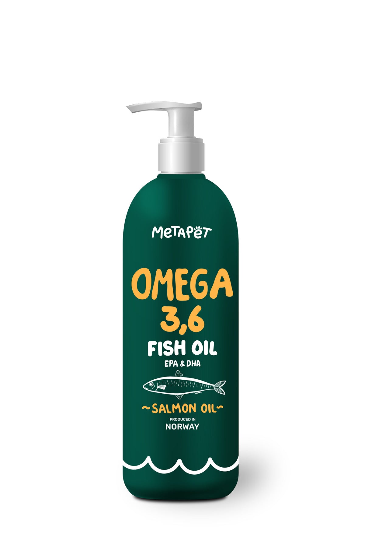
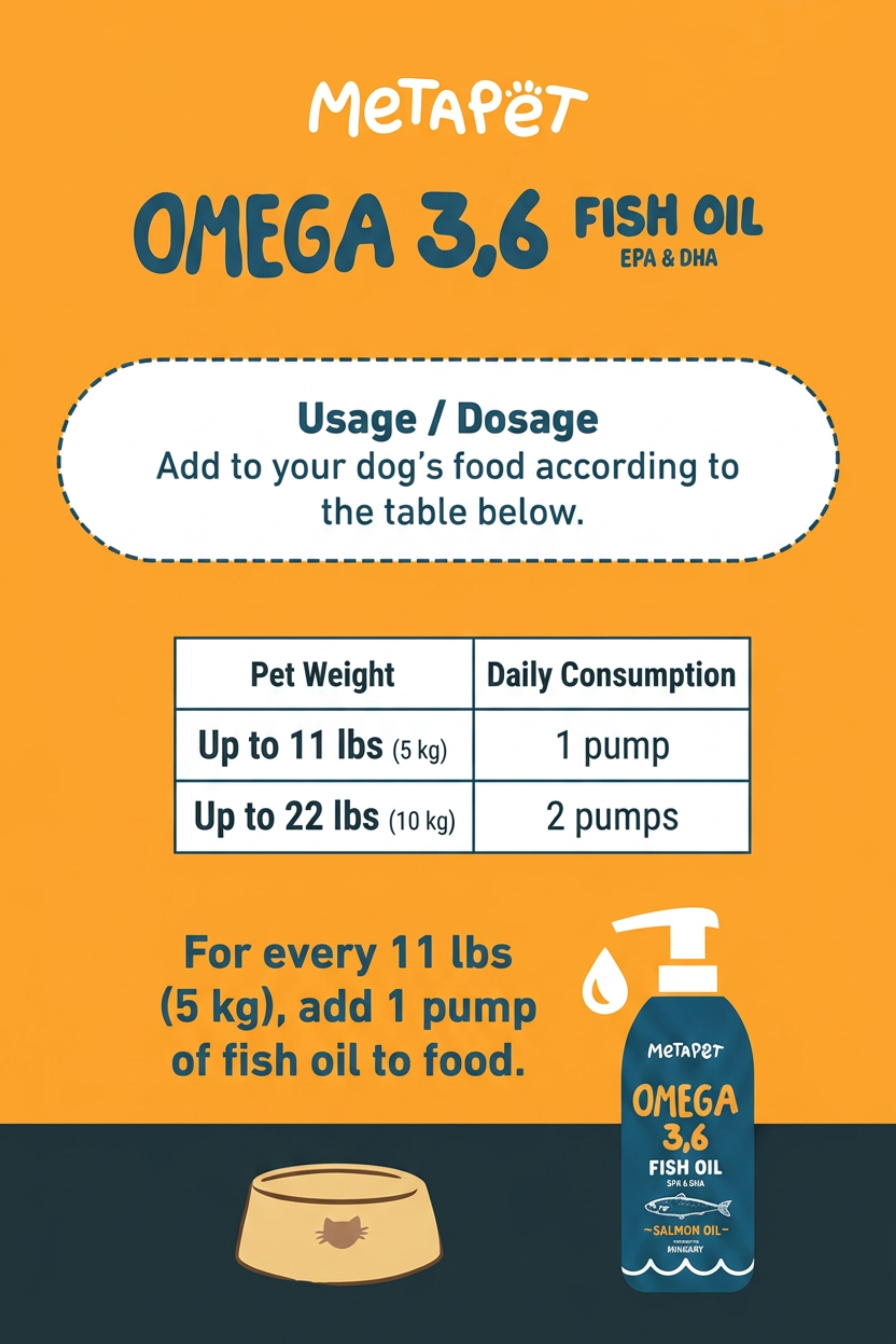
Where a general wellness routine is part of daily life, keep it in proportion. Everyday products such as an omega oil poured over food — for example the MetaPet Omega Salmon Oil for Dogs — are food supplements intended as part of a normal feeding routine. They are a complement to veterinary care rather than a substitute for it, they do not treat or prevent any disease, and in a dog with gallbladder disease or one on a prescribed low-fat diet you should check with your veterinarian before adding any oil or supplement to meals.
Keep a short written log: appetite, energy, any vomiting, stool quality, and gum colour checked weekly in good light. This makes recheck appointments considerably more useful and helps you notice a gradual decline that day-to-day familiarity tends to hide.
Myths and Facts
- Myth: sludge on an ultrasound means gallbladder disease. Sludge is usually an incidental finding of no clinical importance.
- Myth: a mucocele can be dissolved with the right supplement. No over-the-counter product treats a mucocele; management is veterinary.
- Myth: surgery should wait until the dog is actually sick. Outcomes are markedly better with planned surgery than emergency surgery after rupture.
- Myth: removing the gallbladder cripples digestion. Dogs adapt well and live normal lives without one.
- Myth: normal appetite means the gallbladder is fine. Many dogs with mucoceles eat normally until late in the process.
- Fact: breed matters. Shetland Sheepdogs and Cocker Spaniels are notably over-represented.
- Fact: jaundice is always urgent. Yellow gums or eyes in a dog need same-day veterinary assessment.
When to Contact Your Veterinarian
Gallbladder disease produces both slow-burning vague signs and sudden dramatic ones, and both categories deserve a response.
- Book an appointment for: intermittent vomiting, reduced appetite over several days, unexplained lethargy, or a dog who simply seems off.
- Call the same day for: yellowing of the gums, eyes, or skin, persistent vomiting, or abdominal discomfort.
- Seek emergency care for: sudden collapse, severe abdominal pain, a distended painful abdomen, pale gums, or rapid deterioration.
For any medical emergency, contact your veterinarian or your nearest emergency veterinary hospital immediately. A ruptured gallbladder is a surgical emergency where time genuinely changes the outcome.
The Practical Takeaway
The most useful thing an owner can take from all of this is that the two common gallbladder findings sit at opposite ends of the seriousness scale. Sludge on an ultrasound in an otherwise well dog is usually a note in the file. A mucocele is a condition with a real risk of progressing to an emergency.
If your dog belongs to a predisposed breed — Shetland Sheepdog, Cocker Spaniel, Miniature Schnauzer among them — it is worth knowing this exists, and worth taking mildly raised liver values on a routine blood panel seriously enough to follow up rather than repeat in six months and hope.
Where a mucocele is diagnosed, the decisions belong to you and your veterinarian together, and they turn on how advanced it is and how well your dog is. The one principle that holds across nearly every case is that acting from a position of stability beats reacting from a position of crisis. Keep the rechecks, feed the diet properly, and check the gum colour now and then.